O&G
-
Reporting in Adolescent Gynaecology
Dr Joanne Ludlow
Dr Karen Atkin -
Non-lethal Skeletal Dysplasia - A Case Series
-
Non-Invasive Prenatal Screening
-
Endometriosis - A case report
-
O&G Interesting Cases
-
IUGR vs SGA
-
Fetal Neurosonogram
-
Improving the Detection of Cardiac Anomalies Antenatally
-
NIPT vs NT
-
Live scan: Endometriosis (ISUOG) - Canon Medical sponsor
Live scanning for endometriosis (ISUOG) in a methodical way following the IDEA protocol.
-
Can MRI add value to ultrasound in the assessment of female fertility?
-
Polycystic Ovarian Syndrome
-
Pelvic Pain: Besides uterus and ovaries, where else should I be looking?
-
Fertility and Pregnancy Options for Transgender and Gender Non-Conforming People
-
Diagnostic approach to prenatally detected gender anomalies
-
How to perform a gynaecological ultrasound in paediatric or adolescent patient
-
Pregnancy after Caesarean Section
Pregnancy after Caesarean Section. What the sonographer needs to know.
-
Fetal Brain MRI
-
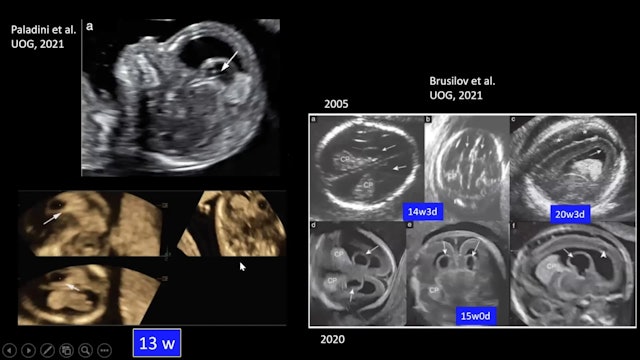
Targeted Neurosonography
-
The fetal brain as demonstrated during the routine second trimester ultrasound
The fetal brain as demonstrated during the routine second trimester ultrasound exam. What can we miss?
-
First Trimester Screening for Fetal Growth Restriction
-
Solid Ovarian Masses: Lessons and Pitfalls
-
Cortical Maldevelopment
-
Familial cancers BRCA, Lynch Syndrome and their associations


